skull anatomy notes pdf
Understanding the human skull is fundamental in numerous fields, and readily available skull anatomy notes in PDF format provide accessible learning resources․ These documents detail cranial and facial structures․
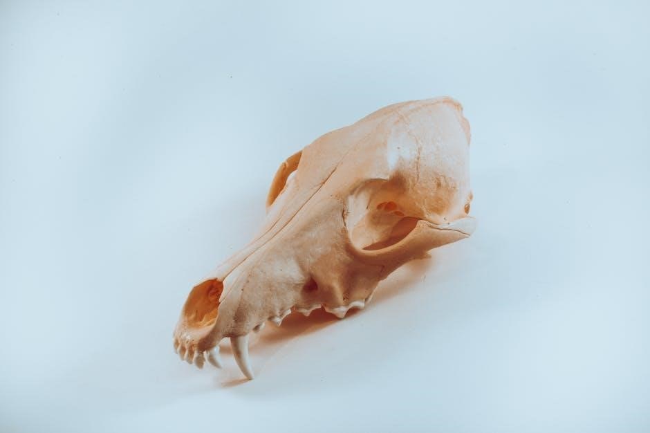
The skull’s primary function is protecting the brain, supporting facial structures, and providing attachment points for muscles․ Detailed diagrams and illustrations enhance comprehension․
Numerous online platforms, like iStock and Adobe Stock, offer a vast collection of labeled skull diagrams and illustrations, often available as downloadable PDFs for study․
A․ Importance of Studying the Skull
The study of skull anatomy is paramount across diverse disciplines, ranging from medicine and anthropology to forensic science and art․ A solid grasp of cranial structures, facilitated by resources like skull anatomy notes in PDF format, is crucial for accurate diagnosis and treatment in medical fields․ Understanding the intricate details of the skull allows healthcare professionals to effectively interpret medical imaging, perform surgical procedures, and diagnose conditions affecting the head and brain․
Furthermore, in forensic science, skeletal remains often provide the primary evidence in identifying individuals and determining the cause of death․ Detailed knowledge of skull morphology, readily available through comprehensive PDF guides, is essential for accurate age estimation, sex determination, and ancestry assessment․ Anthropologists rely on skull analysis to trace human evolution and understand population variations․
Artists, particularly sculptors and illustrators, benefit immensely from a thorough understanding of skull anatomy to accurately depict the human head․ The availability of high-quality, labeled diagrams and illustrations, often compiled in convenient PDF documents, provides invaluable reference material․ Access to these resources, found on platforms like iStock and Adobe Stock, empowers students and professionals alike to deepen their knowledge and refine their skills․
B․ Overview of Skull Function
The human skull performs a multitude of vital functions, extending far beyond simply protecting the brain․ Its robust structure safeguards this delicate organ from physical trauma, while simultaneously providing attachment points for the muscles of the head and neck, enabling a wide range of movements and expressions․ Detailed skull anatomy notes in PDF format often highlight these crucial muscular connections․
Beyond protection and movement, the skull houses the sensory organs – eyes, ears, nose, and mouth – and plays a critical role in facial expression and communication․ The sinuses within the skull contribute to voice resonance and regulate temperature․ Understanding these multifaceted functions is greatly aided by visual resources, such as those available on Adobe Stock and iStock․
Moreover, the skull’s foramina – openings for nerves and blood vessels – are essential for connecting the brain to the rest of the body, facilitating sensory input and motor output․ Comprehensive PDF guides often include detailed diagrams illustrating these pathways․ Studying these functions, supported by accessible resources, is fundamental to appreciating the skull’s complex role in overall human physiology․
C․ Types of Skull Diagrams & Resources (PDF Focus)
A wealth of skull diagrams and resources are available, with skull anatomy notes in PDF format being particularly convenient for study․ These range from basic, labeled illustrations identifying major bones to highly detailed anatomical charts showcasing foramina, processes, and muscle attachments․ Platforms like iStock and Adobe Stock offer extensive collections of these visual aids․
PDF resources often categorize diagrams by view – lateral, anterior, posterior, superior, and inferior – providing a comprehensive understanding of the skull’s three-dimensional structure․ Some PDFs focus on specific aspects, such as cranial nerves or the facial skeleton, offering specialized detail․ Video resources, also found on Adobe Stock, complement static diagrams, demonstrating spatial relationships․
Furthermore, educational institutions and online learning platforms frequently provide downloadable PDF study guides․ These often include quizzes and self-assessment tools․ When selecting resources, consider the level of detail, clarity of labeling, and accuracy of information․ Utilizing a variety of diagram types and PDF guides enhances learning and retention․

II․ Skull Bones: Cranial Vault
The cranial vault, detailed in skull anatomy notes PDF guides, comprises the frontal, parietal, temporal, and occipital bones․ These bones protect the brain effectively․
Understanding their articulations and features is crucial for anatomical study, as illustrated in available diagrams and resources․
A․ Frontal Bone – Anatomy & Features
The frontal bone, extensively covered in skull anatomy notes PDF resources, forms the anterior portion of the cranium and contributes to the orbits․ Its complex structure includes several key features essential for identification and understanding its function․
Anatomically, the frontal bone consists of a squamous part, forming the forehead, and a vertical part contributing to the orbital rims․ The supraorbital margin, a prominent ridge, houses the supraorbital foramen, a pathway for nerves and vessels supplying the forehead and upper eyelid․
Internally, the frontal bone contains the frontal sinuses, air-filled spaces that reduce skull weight and contribute to voice resonance; These sinuses are clinically significant, as inflammation can lead to sinusitis․ The sagittal suture marks the midline articulation with the parietal bones, while the coronal suture connects it to the parietal bones laterally․
Detailed skull anatomy notes PDF often include illustrations highlighting the glabella, the smooth area between the eyebrows, and the brow ridges, providing attachment points for facial muscles․ Understanding these features is vital for medical professionals and students alike, aiding in accurate diagnosis and treatment related to head injuries or facial reconstruction․
Resources like those found on Adobe Stock and iStock provide visual aids complementing textual information, enhancing the learning experience․
B․ Parietal Bones – Structure & Articulations
Parietal bones, thoroughly detailed in skull anatomy notes PDF guides, form the majority of the cranial vault’s superior and lateral walls․ Typically, individuals possess two parietal bones, contributing significantly to the skull’s overall shape and protective function․
Structurally, each parietal bone is roughly quadrilateral, exhibiting a convex external surface and a concave internal surface accommodating the brain․ Key features include the superior and inferior temporal lines, serving as attachment points for muscles involved in mastication and head movement․
Articulations are crucial to understanding parietal bone function․ They connect with the frontal bone via the coronal suture, the occipital bone through the lambdoid suture, and each other along the sagittal suture․ These sutures are fibrous joints, allowing limited movement during infancy but becoming firmly fused in adulthood․
Skull anatomy notes PDF often emphasize the parietal foramen, a small opening near the superior border, allowing passage for emissary veins connecting scalp veins with intracranial venous sinuses․ Visual resources from platforms like Adobe Stock and iStock aid in identifying these features․
Accurate identification of these structures is essential for neurosurgical planning and interpreting skull imaging․
C․ Temporal Bones – Detailed Examination
Temporal bones, comprehensively covered in skull anatomy notes PDF resources, are complex structures forming the lateral and inferior aspects of the cranium․ Located on each side of the head, they house vital structures for hearing and balance․
Detailed examination reveals several key components: the squamous, petrous, mastoid, and tympanic portions․ The petrous portion, particularly, is significant, containing the inner ear and associated structures․ These notes often include labeled diagrams from sources like iStock and Adobe Stock․
The mastoid process, a prominent projection posterior to the ear, serves as an attachment point for neck muscles․ The styloid process, another important feature, provides attachment for ligaments and muscles of the tongue and pharynx․
Numerous foramina perforate the temporal bone, facilitating the passage of cranial nerves (VII, VIII, IX, X) and blood vessels․ Understanding these pathways is crucial, as detailed in skull anatomy notes PDF guides․
Visual aids within these PDFs highlight the intricate anatomy, aiding in the identification of these critical structures and their clinical relevance․
D․ Occipital Bone – Foramen Magnum & More
The occipital bone, thoroughly detailed in skull anatomy notes PDF materials, forms the posterior and inferior portion of the cranium․ Its most prominent feature is the foramen magnum, a large opening through which the spinal cord connects to the brainstem․
These notes, often incorporating illustrations from resources like Adobe Stock and iStock, emphasize the bone’s complex structure․ The occipital bone articulates with the parietal bones via the lambdoid suture, and with the temporal bones forming part of the base of the skull․
External occipital protuberance, a noticeable bump on the posterior surface, serves as an attachment point for ligaments and muscles of the neck․ Superior and inferior nuchal lines radiate laterally from this protuberance․
Several foramina, including the hypoglossal canal (for cranial nerve XII) and the occipital condylar foramina, are present, allowing passage for nerves and vessels․ Skull anatomy notes PDF guides clearly label these structures․
Detailed diagrams within these resources aid in visualizing the bone’s intricate anatomy and understanding its functional significance․

III․ Skull Bones: Facial Skeleton
Facial bones, detailed in skull anatomy notes PDF, form the framework of the face․ These notes illustrate the nasal, maxilla, zygomatic, and mandible bones’ structure and function․
Resources like Adobe Stock provide visual aids for understanding these complex structures, enhancing learning and anatomical comprehension․
A․ Nasal Bones – Form & Function
The nasal bones, frequently detailed within skull anatomy notes PDF resources, are two small, rectangular bones forming the bridge of the nose․ These bones articulate with the frontal bone superiorly and the maxilla laterally, contributing to the overall nasal structure․ Their primary function is to support the cartilage and soft tissues of the external nose, providing shape and protection․
Detailed anatomical illustrations, often found in these PDF guides, showcase the paired nature of the nasal bones and their precise articulation points․ Understanding their form is crucial for comprehending facial aesthetics and potential fracture patterns․ The notes typically highlight the superficial location of these bones, making them susceptible to injury․
Resources like those available on Adobe Stock and iStock offer visual representations of the nasal bones within the context of the entire facial skeleton․ These images, often included in comprehensive skull anatomy PDFs, aid in spatial understanding․ Furthermore, the notes often explain how the nasal bones contribute to the nasal cavity, facilitating olfaction and respiration․ Studying these resources provides a solid foundation for medical professionals and students alike․
The precise descriptions within these PDFs are invaluable for identifying anatomical landmarks and understanding the clinical implications of nasal bone fractures or deformities․
B․ Maxilla – Key Features & Sinuses
The maxilla, extensively covered in skull anatomy notes PDF guides, forms the upper jaw and a significant portion of the facial structure․ Paired bones, they articulate with numerous other cranial bones, including the nasal, zygomatic, and palatine bones․ Key features include the alveolar processes, which house the upper teeth, and the infraorbital foramen, a pathway for nerves and vessels;
Crucially, the maxilla contains the maxillary sinuses – large, air-filled spaces that reduce skull weight and contribute to voice resonance․ Skull anatomy notes PDF resources often dedicate sections to detailing the location, size, and clinical significance of these sinuses, including their susceptibility to infection (sinusitis)․
Visual aids from platforms like Adobe Stock and iStock, frequently incorporated into these PDFs, illustrate the complex anatomy of the maxilla, highlighting the sinus cavities and foramina; These resources demonstrate the maxilla’s role in forming the nasal cavity floor, the orbit’s inferior wall, and the hard palate․
Understanding the maxilla’s intricate structure, as detailed in these notes, is vital for dental procedures, facial reconstruction, and diagnosing sinus-related pathologies․
C․ Zygomatic Bones – Cheekbone Anatomy
Zygomatic bones, commonly known as cheekbones, are prominently featured in skull anatomy notes PDF materials․ These paired bones articulate with the frontal, temporal, maxilla, and sphenoid bones, contributing to the lateral and inferior aspects of the orbit (eye socket)․ Detailed diagrams within these PDFs showcase their quadrangle shape and key landmarks․
A crucial feature is the zygomatic arch, formed by the zygomatic bone and the temporal bone’s zygomatic process․ This arch provides attachment points for the masseter muscle, vital for chewing․ Skull anatomy notes PDF often emphasize the zygomatic bone’s role in facial aesthetics and its importance in facial reconstruction procedures․
Resources like Adobe Stock and iStock provide visual representations, frequently included in these PDFs, illustrating the zygomatic bone’s complex articulation and its contribution to facial contours․ These images highlight the temporal and orbital surfaces, as well as the zygomaticofacial and zygomaticotemporal foramina․
Comprehensive understanding of zygomatic bone anatomy, as presented in these notes, is essential for surgeons, dentists, and those studying craniofacial anatomy․
D․ Mandible – Lower Jaw & its Components
The mandible, or lower jaw, is uniquely the only movable bone of the skull, and its detailed anatomy is thoroughly covered in skull anatomy notes PDF resources․ These PDFs illustrate the mandible’s horseshoe shape, comprised of a body and two rami․ The body forms the chin and provides attachment for muscles involved in chewing and facial expression․
Key components highlighted in these notes include the mental foramen, a crucial landmark for nerve and blood vessel passage, and the alveolar process, which houses the lower teeth․ The rami ascend to articulate with the temporal bone, forming the temporomandibular joint (TMJ)․
Visual aids, often sourced from platforms like Adobe Stock and iStock and included in these PDFs, demonstrate the mandible’s intricate features, such as the coronoid and condylar processes․ These processes serve as attachment points for muscles and contribute to TMJ function․
Understanding the mandible’s anatomy, as detailed in these notes, is critical for dental professionals, surgeons, and anyone studying head and neck anatomy․

IV․ Skull Features: Foramina & Processes
Skull anatomy notes PDF resources detail foramina – openings for nerves and vessels – and processes, bony projections for muscle attachment․ These features are crucial for understanding skull function․
Detailed diagrams illustrate the mastoid and styloid processes, alongside cranial foramina locations, aiding comprehensive anatomical study․
A․ Cranial Foramina – Pathways for Nerves & Vessels
Cranial foramina, meticulously detailed in skull anatomy notes PDF guides, are vital openings within the skull bones, serving as crucial pathways for nerves and blood vessels connecting the brain to the periphery․ These openings aren’t simply holes; their size and shape are specifically adapted to accommodate the structures passing through them, ensuring protection and efficient function․
For example, the foramen magnum, a prominent feature, allows the spinal cord to connect to the brainstem․ Other significant foramina include the optic canal (for the optic nerve), the superior orbital fissure (for cranial nerves and vessels supplying the orbit), and the jugular foramen (for the internal jugular vein and several cranial nerves)․
Understanding the precise location and contents of each foramen is essential for neurological assessment and diagnosis․ PDF resources often include labeled diagrams and illustrations that clearly depict these pathways․ Furthermore, clinical notes within these guides highlight the potential consequences of damage or compression to nerves passing through specific foramina, such as vision loss or facial paralysis․ The study of these features is paramount for medical professionals and students alike․
B․ Mastoid Process – Anatomy & Clinical Significance
The mastoid process, a prominent bony projection located behind the ear, is thoroughly illustrated and explained in detailed skull anatomy notes PDF resources․ This structure, part of the temporal bone, serves as an attachment point for several neck muscles, contributing to head movement and stability․ Its internal structure contains air cells, known as the mastoid air cells, which are interconnected with the middle ear cavity․
Clinically, the mastoid process is significant due to its susceptibility to infection, leading to mastoiditis․ PDF guides often include information on the clinical presentation of mastoiditis, including tenderness, swelling, and potential complications like hearing loss or meningitis․
Furthermore, the mastoid process serves as a landmark for surgical procedures, such as tympanomastoid surgery, aimed at addressing chronic ear infections or cholesteatoma․ Detailed anatomical diagrams within these PDF resources are invaluable for surgeons․ Understanding the intricate anatomy of the mastoid process, including its air cell system, is crucial for accurate diagnosis and effective treatment of related conditions․
C․ Styloid Process – Attachment Points
The styloid process, a slender, pointed projection extending from the inferior surface of the temporal bone, is comprehensively detailed in skull anatomy notes PDF materials․ Though small, this structure serves as a crucial attachment site for several ligaments and muscles, playing a vital role in the support and movement of the hyoid bone and tongue․
Specifically, ligaments connecting the styloid process to the hyoid bone contribute to the hyoid’s suspension, influencing swallowing and speech․ Muscles attaching here include the styloglossus, stylopharyngeus, and stylohyoid, each contributing to tongue movement and pharyngeal elevation․ PDF resources often feature labeled diagrams illustrating these attachments․
Elongation of the styloid process, a condition known as Eagle syndrome, can cause chronic throat pain and discomfort․ Skull anatomy notes in PDF format frequently address this clinical condition, detailing its causes, symptoms, and diagnostic imaging․ Understanding the styloid process’s anatomy and associated structures is essential for recognizing and managing this syndrome effectively․

V․ Skull Diagrams & Visual Aids (PDF Content)
Comprehensive skull anatomy notes in PDF format often include detailed, labeled diagrams․ These visual aids showcase cranial and facial bone structures, enhancing understanding and retention of complex anatomical details․
Illustrations range from artistic renderings to precise anatomical depictions, aiding in spatial visualization․ Many PDFs also feature cranial nerve diagrams for functional study․
A․ Labeled Skull Diagrams – Comprehensive Views
Labeled skull diagrams, frequently found within skull anatomy notes in PDF form, are invaluable tools for students and professionals alike․ These diagrams provide comprehensive views of the skull, meticulously identifying each bone and its key features․ Resources like iStock and Adobe Stock offer a wealth of these visual aids, often downloadable for offline study․
Superior diagrams typically showcase multiple perspectives – anterior, posterior, lateral, and inferior – allowing for a complete understanding of the skull’s three-dimensional structure․ Each bone, including the frontal, parietal, temporal, occipital, sphenoid, and ethmoid bones of the cranial vault, is clearly labeled․ Similarly, facial bones such as the nasal, maxilla, zygomatic, and mandible are precisely identified․
Beyond simple bone identification, advanced diagrams often highlight important foramina (openings for nerves and vessels) and processes (projections for muscle attachment)․ These details are crucial for understanding the skull’s functional anatomy․ The quality of these diagrams varies, with premium resources offering higher resolution and greater detail․ Utilizing these PDFs effectively requires consistent review and correlation with anatomical models or real specimens when possible․
Furthermore, color-coding is frequently employed to differentiate between individual bones or to emphasize specific anatomical landmarks, improving clarity and memorization․ The availability of these resources in PDF format ensures easy access and portability for convenient study․
B․ Skull Anatomy Illustrations – Artistic Representations
While labeled diagrams within skull anatomy notes in PDFs prioritize accuracy, artistic illustrations offer a complementary approach to understanding the skull’s complex structure․ These representations, readily available on platforms like iStock and Adobe Stock, often emphasize form and spatial relationships in a visually engaging manner․

Unlike strictly anatomical diagrams, illustrations may employ shading, color, and artistic license to highlight specific features or to create a more aesthetically pleasing image․ This can be particularly helpful for visualizing the skull’s contours and appreciating its overall shape․ Many illustrations depict the skull in various artistic styles, ranging from realistic renderings to stylized depictions․
These artistic interpretations can aid in memorization by providing a different perspective on the anatomy․ They can also be useful for understanding how the skull relates to the surrounding soft tissues․ However, it’s crucial to remember that illustrations are not always perfectly accurate and should be used in conjunction with precise anatomical diagrams․

PDF collections often include a mix of both diagrammatic and illustrative content, providing a well-rounded learning experience․ The availability of these resources in digital format allows for easy zooming and detailed examination of the artwork․
C․ Cranial Nerves Diagram – Sensory & Motor Functions
Comprehensive skull anatomy notes in PDF format invariably include detailed diagrams illustrating the cranial nerves – twelve pairs responsible for sensory and motor functions of the head and neck․ These diagrams are crucial for understanding the neurological connections within the skull․
These diagrams typically depict the origin and pathway of each nerve, clearly labeling its name, number, and primary functions․ Sensory nerves transmit information to the brain, while motor nerves carry signals from the brain to muscles and glands․ Some cranial nerves have both sensory and motor components․
Resources like those found on iStock showcase cranial nerves paired with anatomical sensory functions in outline diagrams․ These visual aids help students correlate specific nerve pathways with their corresponding sensory or motor effects․ Understanding these connections is vital for diagnosing neurological disorders․
PDF versions allow for close examination of the intricate nerve pathways․ The diagrams often include information about the foramen through which each nerve exits the skull, further enhancing anatomical understanding․ Studying these diagrams alongside skull bone illustrations provides a holistic view․